Webinar #5: 9 December 2021 at 11:00 AM ET (UTC -4:00)
A Look at the History of Internationally Recognized Colombian Time Scale UTC (INM) Optical Clocks with Trapped Ions
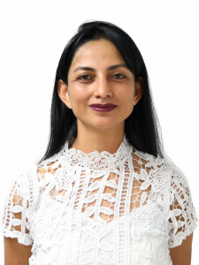
Liz Forero
Liz C. Hernández Forero is Electronic Engineer from Universidad Distrital Francisco José de Caldas and MSc. in Electronic and Computer Engineering - Biomedical Engineering subarea - from Universidad de los Andes, both in Colombia. She has knowledge and experience in the area of control and automation, experience in metrology since May 2008, and head of Time and Frequency Laboratory at National Metrology Institute of Colombia since 2014. Guest Scientist in the Time and Frequency Division at Atomic Standards Group of National Institute of Standards and Technology (NIST) at Boulder, Colorado, United States. She was elected as Vice-Chair of the Time and Frequency Metrology Working Group (TFMWG) of the Inter American Metrology System (SIM - Sistema Interamericano de Metrología) since July 2021. Her interest is growing as a scientist in the Time and Frequency metrology area contributing to national and international cooperation in science and technology, as well as improving her pedagogical skills for the transfer of knowledge in metrology.
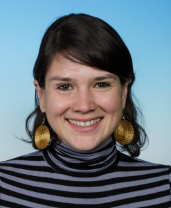
Milena Guevara Bertsch
Milena Guevara Bertsch did her Bachelor's and Master's studies at the University of Costa Rica. During her Master's program, she worked at the Centro de Investigación en Ciencia e Ingeniería de Materiales in the implementation of trapped atoms in optical lattices for the study of surface layers. She worked in the construction of a Zeeman slower and the implementation of Low Energy Electron Diffraction to study the adsorption of water molecules on magnesium oxide surfaces. She is currently doing her Ph.D. studies in the Quantum Optics and Spectroscopy group under the supervision of Rainer Blatt and Christian Roos at the Institute for Quantum Optics and Quantum Information (IQOQI) in Innsbruck, Austria. She is focused on the development of optical clocks with trapped calcium and aluminum ions.
Talk #1
A Look at the History of Internationally Recognized Colombian Time Scale UTC (INM)
Just as every country has its own national anthem, every country designates which entity in the national territory provides the time used for activities associated with legal compliance. That time in the scientific field is called “time scale”. This talk will discuss how the Colombian time scale was developed, how it is generated, and what it is used for in Colombia. Legal Time is important for the academic, scientific, and industrial groups, as well as the citizens. Each group needs different levels of accuracy depending on the intended use of their event dating activities. Therefore, the achievements in establishing the Calibration and Measurement Capabilities (CMC) for the magnitude of time and frequency in Colombia over the last 13 years are presented and how these achievements are reflected in the dissemination of metrology in Colombia, being the first CMC published in the Key Comparison Database (KCDB) of the BIPM (for its French acronym, Bureau International des Poids et Mesures) in the country’s history. The talk will show that international cooperation has been a key tool for the current development of the Time and Frequency Laboratory of the National Metrology Institute of Colombia (INM) and to expand the dissemination of metrological traceability to the International System (SI) of units in the country together with proposals of further work.
Talk #2
Optical Clocks with Trapped Ions
“Never measure anything but frequency” was the motto of Arthur Schawallow. Through the measurement of frequency, we can measure any other quantity and inherit, in the process, the same exactitude. By the careful determination of the perturbation of the frequency of a given oscillator, we can fully characterize its environment: the electric and magnetic fields or the mass of the particles that surround it and even observe relativistic effects. Our understanding of nature seems to be limited by our capacity to measure frequency. What are the key ingredients to building the optimal oscillator? How can we reach and break the limits of precision and accuracy?
This brief talk introduces the development of optical clocks as possible answers to these questions. Optical clocks are based on the precise and accurate interrogation of atomic transitions. These kinds of clocks work in the optical regime of the electromagnetic spectrum, meaning that they operate at transitions that oscillate at more than 400 THz with bandwidths on the sub-Hertz level, allowing us to reach accuracies up to one part in 1018 or even lower. During this talk, I will introduce the necessary elements for the development of optical clocks with trapped calcium and aluminum ions.
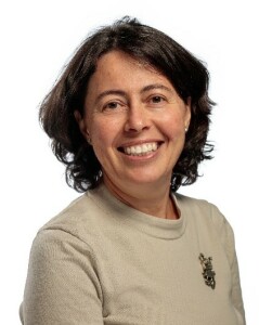
Marina Gertsvolf
Moderator
Dr. Marina Gertsvolf is the Team Leader for Frequency and Time (F&T) group at NRC and is responsible for realizing the second, an SI unit of time, and for maintaining and disseminating the official time for Canada, UTC(NRC).
Dr. Gertsvolf received her Ph.D. from the University of Ottawa in 2009 and joined NRC as a research officer the same year. She has been working on a variety of projects from Caesium Fountain development to frequency comb construction, timescale generation, and maintenance, as well as on calibrations and traceability. Dr. Gertsvolf serves on several international committees and working groups. Among others, she is the Commission Chair of Canadian National Committee for the International Union of Radio Science (URSI), the Chair of the International Atomic Time Working Group at the Consultative Committee of Time and Frequency (CCTF-WGTAI) and the Deputy Technical Chair of the Systema Interamericano de Metrologia (SIM).
In 2016 Dr. Gertsvolf became the F&T team leader and has been leading the group and the development of the next generation frequency standards and dissemination services that meet and exceed current industry and society needs. NRC F&T group operates and develops among others, the caesium fountain atomic clock, the primary realization of SI second; the single trapped strontium ion clock, the most accurate frequency standard in Canada and one of the best in the world; the nano-second accuracy time dissemination service to remote clients in support of the critical infrastructure needs, and the frequency comb systems for frequency calibration and comparisons.
Webinar #4: 27 September 2021 at 2:00 PM EDT (UTC -4:00)
Piloting a Tele-ultrasound Diagnostic System for Deployment in Rural Areas of Peru and RBTU Brazilian Platform for Ultrasound Research
Benjamin Castaneda Aphan
Benjamin Castaneda is a Full professor of Biomedical Engineering and Director of Research & Innovation in the Department of Engineering at the Pontificia Universidad Católica del Perú (PUCP). He obtained his M.Sc. in Computer Engineering from Rochester Institute of Technology, NY, and his Ph.D. in Electrical Engineering from the University of Rochester, NY. After his Ph.D., he founded the Medical Imaging Laboratory at PUCP. He has more than 15 years of experience in biomedical Ultrasound and medical imaging analysis. Dr. Castaneda’s research topics include quantitative elastographic imaging (breast cancer diagnosis, prostate cancer detection, skin characterization), computed aided diagnosis tools (automated diagnosis of Tuberculosis, spondyloarthritis, follow-up of treatment for Leishmaniasis, and preventive diagnosis in maternal-perinatal health), and telemedicine (obstetric ultrasound and colposcopy). For his research work, he has twice been a finalist for the Young Investigator Award from the American Institute of Ultrasound in Medicine (2007, 2011). He obtained an honorable mention from the worldwide engineering contest of Mondialogo (sponsored by UNESCO and Daimler, 2007), obtained an honorable mention in the SPIE Medical Imaging International Conference (2008). In 2013, Dr. Castaneda received the Academic Innovator Award from the Peruvian Government for his continuous work in the development of medical technology (SINACYT/CONCYTEC, 2013). The same year, he won the Best Patent from the Peruvian Government (INDECOPI, 2013) for an automated staining system for tuberculosis detection. The same invention received a silver medal at the International Exhibition of Inventions of Geneva (2014). He is also the founder of Medical Innovation & Technology, a Peruvian start-up focused on the development of telemedicine technology for rural areas. He is currently a member of the Peruvian Committee for Health Informatics and an Associate Member of the IEEE Bio Imaging and Signal Processing Technical Committee.
Talk #1
Piloting a Tele-ultrasound Diagnostic System for Deployment in Rural Areas of Peru
Millions lack access to adequate diagnostic imaging services in rural areas worldwide. As a low-cost and portable imaging modality, ultrasound will play a key role in correcting these disparities. However, access to ultrasound technology is limited by the availability of trained sonographers and readers in rural areas. As a solution, an ultrasound-based telemedicine imaging system has been developed for deployment in rural areas in which ultrasound naïve rural health workers perform volume sweep imaging (VSI) protocols based on only external body landmarks for diagnosing obstetrical, gallbladder, and thyroid pathology. Our telemedicine ultrasound system was piloted in an academic setting at the Pontifical Catholic University of Lima (PUCP) and in the Peruvian rainforest. Health workers were trained on VSI protocols, image capture, labeling, and electronic transmission over 2-3 days. Trainees then obtained images from patients requiring obstetrics, gallbladder, or thyroid scans. Images were uploaded to a cloud-based system and then downloaded and read by radiologists all using custom software. Preliminary results suggest that trainees can learn VSI protocols within 3 days. Our complete telemedicine system can provide a low-cost and scalable diagnostic service suitable for deployment in rural areas.
Eduardo Tavares Costa
Eduardo T. Costa received his BSc degree in Electrical Engineering from the University of São Paulo in 1978, the MSc in Biomedical Engineering from the University of Campinas (1985), and his Ph.D. in Medical Engineering and Physics from the King’s College of the University of London (1989). He is a Full-time Professor at the Department of Electronics and Biomedical Engineering (DEEB) of the School of Electrical and Computer Engineering (FEEC) of the University of Campinas (Unicamp). He is an Associate Researcher of the Center for Biomedical Engineering (CEB) being its director for two terms (2005-2008 and 2008-2011). He served as President of SBEB (Brazilian Society of Biomedical Engineering) for two terms (2002-2004 and 2011-2012). He teaches electronics and biomedical engineering for undergraduate and graduate students including MSc and Ph.D. supervision. He is interested in ultrasound circuitry (with firmware/software) for medical and biological applications and in biomedical instrumentation development.
Amanda Costa Martinez
Amanda C. Martinez received her BSc degree in Electronic Engineering from the Federal Technological University of Paraná in 2016, the MSc in Biomedical Engineering from the University of Campinas (2019), and is currently a Ph.D. candidate in Biomedical Engineering at the University of Campinas. She is interested in ultrasound technology for medical and biological applications and in biomedical instrumentation development.
Talk #2
RBTU Brazilian Platform for Ultrasound Research
The talk will show an overview of the platform for ultrasound research developed in a joint effort of researchers of five Brazilian universities (UNICAMP, USP, UTFPR, UFRJ, UFSCar), one clinical research institute (INCOR-SP), and one technical institute (Eldorado), that formed RBTU – Rede Brasileira de Técnicas de Ultrassom (Brazilian Network for Ultrasound Techniques). The development of the platform was funded by FINEP/MCTI and FNS/MS, Brazilian agencies linked to the Ministry of Science, Technology, and Innovation (MCTI) and the Ministry of Health (MS). The academic team was composed of 15 researchers and MSc and Ph.D. students and the technical institute involved more than 26 hardware and software engineers. By the end of this project, two versions of this ultrasound research platform were developed. In this presentation, we will show the main characteristics of the second and more flexible platform version. It consists of a 64-channel FPGA-based hardware with two 32-channel transmitters (TX) and receiver (RX) boards, a control board, multiplex, and backplane boards. It is possible to use a 128-element matrix transducer using the mux board. The hardware provides all RF data processed in a GPU board inside a personal computer equipped with two 3.0 USB interfaces (each interface is connected to a 32-channel RX board). The user interface allows the researcher to define and produce traditional (B-mode, Doppler, and M-mode) images and, also, create different imaging techniques. It is possible to acquire up to 128 individual RF A-line.
In the RX board is actually implemented the traditional delay-and-sum method before data are sent to be processed in the PC GPU board. Each RBTU research group is now using the platform to conduct their own research. In our group, we are using the platform to implement the fast plane-wave imaging technique and also to characterize ultrasound transducers. All new applications will be made available to RBTU members.
Jean-luc Gennisson
Moderator
Joining the CNRS (the French National Center for Science) in 2005 as a permanent researcher, Gennisson has mainly developed the ultrafast ultrasound imaging technique and especially the elastography technique to quantify the stiffness of biological tissue. Gennisson drove more than 15 clinical protocols on elastography development. This work took the continuation of his Ph.D. which concerned a 1D ultrasound imaging device, the “Fibroscan®” which is now commercialized by the company Echosens® and use for the clinical diagnosis of liver fibrosis. During these years of development, he has contributed to improving the elastography technique by creating new 2D and 3D modes, allowing the quantification of other mechanical parameters of tissues such as viscosity, anisotropy, elastic nonlinearity. In 2014, he has conceptualized and built the first 4D ultrafast ultrasound platform to provide volume information’s in elastography but with no real-time approach. In 2017, Gennisson moved to IR4M laboratory that became BIOMAPS laboratory in 2020, to become head of the team “Methodological developments and instrumentation” (25 people with permanent position).
Webinar #3: 30 August, 2021 at 2:00pm EDT (UTC -4:00)
Featured Topic: Acoustic Vortex Beams - From Particle Levitation in Air to Cell Manipulation
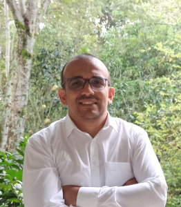
Joao Luis Ealo Cuello
Joao Ealo is professor at the School of Mechanical Engineering of the Universidad del Valle, Cali, Colombia, since 2002. He received the B.Sc. in Mechanical Engineering from the University of Ibagué, Colombia, in 1998, and the M.Sc. in Industrial Control Systems from the University of Valladolid, Spain, in 2000. In 2009, He obtained a doctorate degree in Mechanical Engineering from the Polytechnic University of Madrid, Madrid, Spain, supported by the Institute for Industrial Automation of the Spanish Research Council (CSIC) and the Universidad del Valle. In 2010 founded the Laboratory in Vibrations and Acoustics (LaVA), through which he conducts research and development activities along with his students and collaborators. Current research interests at LaVA are aimed to: a) exploring different transducers technologies to be used in air-coupled ultrasonic applications, such as acoustic imaging, non-destructive testing, robot navigation, particle manipulation, etc.; b) material characterization through non-contact ultrasonic techniques, this includes laser-based ultrasound and ultrasonic spectroscopy; c) modeling, fabrication and characterization of electromechanical-acoustic sources;d) Vibration and acoustics in industrial environments; d) vibroacoustic characterization of Colombian autochthonous musical instruments and e) acoustic vortex beams.
Main Talk
Electroactive Diffraction Gratings: An Efficient Alternative for High-Quality Airborne Ultrasound Vortex Beam Generation
Acoustic vortex beams (AVB) have shown great potential in different emerging applications, such as contactless particle manipulation, transfer of angular momentum to matter, acoustic imaging, communications, and more recently, in biomedical applications. Different methods for AVB generation have been proposed, e.g., physically spiral sources, phase-driven sources, passive metastructured devices and electroactive spiral gratings. Notably, most of them have been developed in water, whereas airborne technologies offer interesting advantages, such as the free access to the manipulated sample.
In this work an efficient technique to easily generate high-quality structured acoustic beams in air is presented. This new approach eliminates reflection losses included in previously reported passive approaches that combine an acoustic source with a diffraction grating. This is possible by converting the grating into an emitter. To demonstrate the versatility of this approach, active spiral-shaped diffraction gratings were fabricated to generate AVB over a broad range of frequencies, i.e., from 70 kHz to beyond 300 kHz, in air. By varying the excitation frequency, a fine and continuous tuning of the focal length of the resultant AVB can be achieved, i.e., the beam can be axially steered while their spatial distribution is preserved. Experimental results from two grating prototypes are included: an Archimedean spiral able to generate simultaneous higher order Bessel beams with different topological charges along the propagation axis and a spiral-Fresnel Zone Plate that allows for sharply focused AVG. The experiments show a good agreement with simulations. The versatility and simplicity of the fabrication process make this technique highly suitable for emerging applications such as particle manipulation, imaging, and transfer of angular momentum to matter. A comparison between the different fabrication methods reported for AVB generation is included in this presentation.
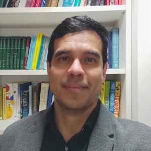
Glauber Silva
Glauber T. Silva (CNPq research fellow) received his Ph.D. in nonlinear ultrasonic imaging from the Federal University of Pernambuco/Mayo Clinic (2003). After postdoctoral positions at Mayo Clinic College of Medicine and Institute of Pure and Applied Mathematics (2007), he joined the Institute of Physics at the Federal University of Alagoas (2008), where he is presently an associate professor. He was a visiting scholar at the Technical University of Denmark (2013), an associate researcher at the University of Bristol (Royal Society, 2017-2019), and a visiting researcher (Chair Total) at ESPCI ParisTech (2019). He is an associate editor of IEEE Transactions on Ultrasonic Ferroelectric and Frequency Control and Applied Acoustics. His current research interests include microparticle and cell manipulation in microfluidics and ultrasonic imaging.
Talk #2
3D Printed Acoustofluidic Devices for Biospectroscopy Applications
Acoustofluidic devices combine the forces and torques produced by ultrasonic waves with microfluidics technology to separate, enrich, pattern, and rotate cells, bacteria, and other microorganisms. In this talk, I will discuss the physical principles of 3D printed acoustofluidic devices used for cell levitation and aggregation. The cellular aggregate is formed in a controlled microenvironment at hundreds of micrometers above the device substrate. This is particularly suitable for Raman biospectroscopy, where signal interferences from the substrate are avoided. In turn, the Raman spectrum reveals the chemical bonds present in a cell. Also, the aggregation stability allows for individual cell Raman-spectrum acquisition. Acoustofluidic-assisted biospectroscopy of murine macrophages and red blood cells is illustrated with preferred quality over ordinary Raman strategies.
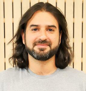
David Espindola
Moderator
Dr. David Espíndola was born in Chillán, Chile, in 1986. He received the Ph.D. degree in physics from the Universidad de Santiago de Chile, Santiago, Chile, in 2012. As part of his Ph.D. dissertation, he studied the interaction wave-particle in granular materials. He pursued post-doctoral research at the Institut d’Alembert, Sorbonne Université, Paris, France, where he started conducting research on medical ultrasound. He also held a post-doctoral position with The University of North Carolina at Chapel Hill, Chapel Hill, NC, USA, where he also was a Research Assistant Professor. He is currently an Associate Professor at the Instituto de Ciencias de la Ingeniería at the Universidad de O’Higgins in Chile. His research interests are the linear and nonlinear elastic wave propagation in soft materials, ultrasound super-resolution imaging and the elasto-acoustics in complex medium.
Webinar #2: 28 June, 2021 at 2:00pm EDT (UTC -4:00)
Featured Topic: Quantitative Ultrasound and High Frame Rate Volume Imaging
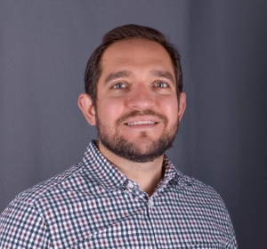
Ivan Miguel Rosado Mendez, Ph.D.
Ivan M. Rosado-Mendez received his B.S. degree in engineering physics from the Tecnologico de Monterrey, his M.S. degree in medical physics from the National Autonomous University of Mexico, and his Ph.D. degree in medical physics from the University of Wisconsin–Madison. After graduating in 2014, he stayed at UW-Madison as a postdoctoral fellow and then as a scientist. From 2017 to 2021, he was an Assistant Professor at the Institute of Physics of the National Autonomous University of Mexico. Dr. Rosado-Mendez recently returned to UW-Madison to join the Quantitative Ultrasound Laboratory as an assistant professor. His research interests include quantification of coherent and incoherent ultrasonic scattering for tissue characterization as well as the analysis of tissue viscoelasticity using shear wave elastography.
Main Talk
Quantitative Ultrasound in Obstetrics and Neonatology
Quantitative Ultrasound (QUS) has the potential to provide noninvasive biomarkers of tissue structure and function in a practical and safe way. This talk will cover applications of two QUS methods, shear wave elasticity imaging and ultrasound backscatter spectroscopy, for the assessment of normal remodeling and damage of structurally complex tissues: the uterine cervix and the neonatal brain. In the cervix, shear wave elasticity imaging is investigated to analyze the frequency dependence of the phase velocity (dispersion) to learn about the structural basis of the cervical remodeling process during pregnancy. In the neonatal brain, ultrasound backscatter spectroscopy is being investigated as a potential tool for detecting cell death caused by different insults, such as long exposures to anesthetics. The talk will conclude with a discussion on the perspective for an extended application of these techniques.
Miguel Bernal
Miguel Bernal received the degree in biomedical engineering from the School of Engineering of Antioquia, Medellin, Colombia. He then obtained his Ph.D. degree in Biomedical Sciences and Biomedical Engineering from Mayo Clinic College of Medicine, Rochester, MN, USA under the supervision of Dr. James F. Greenleaf. He subsequently did a 4-year postdoctoral fellowship at the Institute Langevin in Paris France with Dr. Mickael Tanter. After his time in France, he returned to Colombia and completed a second postdoctoral fellowship at the Universidad Pontificia Bolivariana. In 2018, Dr. Bernal joined Verasonics Inc, in Kirkland WA, USA as an Ultrasound Scientist. Finally, in 2019 Dr. Bernal returned to Colombia as the Senior Ultrasound Scientist of Verasonics SAS, a subsidiary of Verasonics Inc for South America.
Industry Speaker
High Frame Rate Volume Imaging through Sparse Random Aperture Compounding
High frame-rate volume imaging (HFVI) has become an important area of research in the last few years since it provided high temporal resolution necessary for advanced image processing. Unfortunately, HFVI usually requires ultrasound systems with a large channel count to achieve rates in the kHz order. These systems are usually expensive which makes their use limited to a few laboratories around the world. In 2020, Verasonics introduced the concept of Sparse Random Aperture Compounding (SRAC) to help overcome this limitation. SRAC was developed for ultrasound systems with a significantly lower number of channels than elements in a matrix probe. The idea is to perform compounding of random apertures to balance image quality and frame rate. In the current work we studied the impact of random aperture optimization on the image quality and evaluated the compounding of complementary and non-complementary random apertures. While our previous study showed the benefits of using SRAC for HFVI, random aperture optimization is crucial to improve image quality while preserving high frame rates.
Roberto Lavarello
Moderator
Roberto Lavarello received his B.Sc. degree in Electronics Engineering from the Pontificia Universidad Católica del Perú in 2000, and his M.Sc. and Ph.D. degrees in Electrical and Computer Engineering from the University of Illinois at Urbana-Champaign in 2005 and 2009, respectively. He is currently a full professor at the Department of Engineering of the Pontificia Universidad Católica del Perú and the director of the Medical Imaging Laboratory, the M.SC. in Biomedical Engineering, and the Ph.D. in Engineering programs from the same institution. His research is primarily focused on the reconstruction and processing of images for the non-invasive assessment of pathological conditions. He is a senior member of IEEE and a former Fulbright scholarship recipient. He served as an Associate Editor for the IEEE Transactions on Biomedical Engineering (2010-2012) and is currently an Associate Editor for the IEEE Transactions on Ultrasonics, Ferroelectrics and Frequency Control and the IEEE Transactions on Medical Imaging, and an editorial board member for the IEEE Open Access Journal of Engineering in Medicine and Biology. He has served as IEEE EMBS Peru Section Chapter chair (2014-2016) and is currently the R9 representative at the IEEE EMBS AdCom (2017-2022), the chair-of the IEEE Transactions on Medical Imaging Steering Committee, the chair of the the IEEE International Symposium on Biomedical Imaging Steering Committee, the co-chair of the backscatter coefficient group of the the AIUM/QIBA Pulse-Echo Quantitative Ultrasound Biomarker committee. and a member of the IEEE EMBS Technical Committee on Biomedical Imaging and Image Processing, the IEEE SPS Technical Committee on Bio Imaging and Signal Processing, the Technical Program Committee of the IEEE International Ultrasonics Symposium, and the IEEE UFFC Diversity and Inclusion committee.
Webinar #1: 31 May, 2021 at 2:00 PM EDT (UTC-4:00)
Featured Topic: Breast Cancer Ultrasound Imaging
Ana B. Ramirez
Ana B. Ramirez received the B.E.E degree from the Universidad Industrial de Santander (UIS), Colombia; and the Ph.D. degree in Electrical Engineering from University of Delaware, USA. She held a post-doc position in the Delft University of technology University at the Acoustical Wavefield Imaging group. She is currently a Full Professor of the Electrical, Electronics, and Communications Engineering Department at Universidad Industrial de Santander, in Colombia. Her fields of interest include acoustic signal processing applied to medical and geophysical data. Her active fields of research are full-waveform inversion of medical and geophysical data, and compressive sensing, and sparse signal processing. She has published more than 20 papers in the areas of interest. Also, she has led several research projects funded by UIS, Colombian Ministry of Science, Ecopetrol, and the US Research Army Lab.
Main Talk
Comparing Full Waveform Inversion Methods for Ultrasound Imaging for Breast Cancer Detection
Current ultrasound imaging techniques use only part of the information enclosed in the recorded high-frequency sound waves limiting the quality of the information present in the reconstructed image. Advanced ultrasound imaging methods, known as full waveform inversion, use all available information enclosed in the recorded field – including multiple scattering, dispersion, and diffraction effects – to improve the image quality and give accurate quantitative information about the tissue parameters. Full-wave inversion (FWI) may be implemented in either the time or the frequency domain but determining which method should be used is not simple. In this webinar, we will discuss two different non-linear FWI imaging methods: time-domain inversion (TDI) and frequency-domain contrast source inversion (CSI). The methods were tested on the reconstruction of the same synthetic data; a 2-D scan from a circular transducer array enclosing a cancerous breast model. Both methods were evaluated in noise-free and noisy scenarios. Also, the reciprocity of sources and receivers was evaluated as well as the computational complexity of the methods
Carla Silva Perez
Carla Silva Perez received her Ph.D. in health sciences from the Ribeirão Preto School of Medicine at the University of São Paulo (FMRP-USP), Brazil with a collaborative period in the University of Lisbon (Portugal). She also received a Master's degree in Health Sciences from the University of São Paulo; graduation in Physiotherapy from the University of São Paulo and is a member of the research group at the Dermatofunctional Evaluation and Intervention Laboratory (LAIDEF) at FMRP-USP.
Early-career Short Talk
Lymphedema, in Survivors of Breast Cancer
Current ultrasound imaging techniques use only part of the information enclosed in the recorded high-frequency sound waves limiting the quality of the information present in the reconstructed image. Advanced ultrasound imaging methods, known as full waveform inversion, use all available information enclosed in the recorded field – including multiple scattering, dispersion, and diffraction effects – to improve the image quality and give accurate quantitative information about the tissue parameters. Full-wave inversion (FWI) may be implemented in either the time or the frequency domain but determining which method should be used is not simple. In this webinar, we will discuss two different non-linear FWI imaging methods: time-domain inversion (TDI) and frequency-domain contrast source inversion (CSI). The methods were tested on the reconstruction of the same synthetic data; a 2-D scan from a circular transducer array enclosing a cancerous breast model. Both methods were evaluated in noise-free and noisy scenarios. Also, the reciprocity of sources and receivers was evaluated as well as the computational complexity of the methods
Chris de Korte
Moderator
Chris L. de Korte is a Professor of Medical Ultrasound Techniques at Radboud University Medical Center since 2015 and a professor of medical ultrasound imaging at the University of Twente since 2016. He studied electrical engineering at the Eindhoven University of Technology. In 1999, he obtained his Ph.D. at the Biomedical Engineering Group of the Thoraxcentre, Erasmus University Rotterdam on his thesis Intravascular Ultrasound Elastography. In 2002 he joined the Clinical Physics Laboratory, Department of Pediatrics of the Radboud University Nijmegen Medical Center of which he became head in 2004. In 2006 he was registered as Medical Physicist. Since 2012, he is chair of the Medical UltraSound Imaging Centre at the Department of Medical Imaging: Radiology of Radboud University Medical Centre. His research is on functional imaging using Ultrasound with a focus on cardiovascular and oncological applications. For his research, he received multiple grants from the Dutch Technology Foundation (STW) and a VENI (2000), VIDI (2006), and VICI (2011) grant from The Netherlands Organization for Scientific Research (NWO). In 2018, a consortium under his leadership was awarded the NWO-TTW Perspectief grant (4M euros). Prof. de Korte is president of the Netherlands Society for Medical Ultrasound (NVMU) and national delegate of the European Federation of Societies for Ultrasound in Medicine and Biology (EFSUMB). He is an associate editor for IEEE Transactions on UFFC-S and the Journal of Medical Ultrasonics. He serves as an editorial board member of Ultrasound in Medicine and Biology and the Journal of the British Medical Ultrasound Society and is a member of the Technical Program Committee of the IEEE International Ultrasonics Conferences.

